CONGENITAL CLOUDY CORNEA
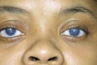
A variety of genetic, metabolic, developmental, and idiopathic causes can result in congenital clouding of the cornea. Note the image below.
 Clouding of the cornea since childhood.
A common reason for congenital clouding of the cornea is congenital glaucoma.
Other major causes of corneal clouding include the following:
The following is a mnemonic for the causes of congenital clouding of the cornea:
Other rarer causes of congenital clouding of the cornea include the following: cornea plana, corneal keloids, oculoauriculovertebral (OAV) dysplasia (Goldenhar-Gorlin syndrome), congenital corneal ectasia, congenital hereditary stromal dystrophy, posterior polymorphous dystrophy, and Fryns syndrome.
Sclerocornea
Sclerocornea is an uncommon developmental abnormality of the anterior segment due to mesenchymal dysgenesis. Sclerocornea manifests as a stationary congenital anomaly. It is usually seen as an isolated ocular abnormality involving both eyes, although it can occur unilaterally. This condition typically occurs sporadically but may also have a familial or autosomal dominant inheritance pattern.
On clinical evaluation, patients with partial sclerocornea have a peripheral, white, vascularized, 1- to 2-mm corneal rim that blends with the sclera, obliterating the limbus. The central cornea is generally normal. In total sclerocornea, the entire cornea is involved, but the center of the cornea is clearer than the periphery. This finding distinguishes it from Peters anomaly, in which the center is most opaque. The opacification affects the full thickness stroma and limits visualization of the posterior corneal surface and of the intraocular structures.
Histopathology reveals disorganized collagenous tissue containing fibrils that is larger than normal. Potential coexisting abnormalities include a shallow anterior chamber, abnormalities of the iris and the lens, and microphthalmos. Systemic abnormalities, such as limb deformities and craniofacial and genitourinary defects, can also accompany this finding. In generalized sclerocornea, early keratoplasty should be considered to provide vision, although the prognosis is guarded.[1]
Descemet membrane tears
Forceps-induced obstetric trauma, with resultant Descemet membrane tears and corneal edema and clouding, is a cause of corneal clouding. This clouding is differentiated from primary congenital glaucoma (PCG) by the presence of periorbital soft tissue trauma, normal intraocular pressure (IOP), and the frequently vertical orientation of the Descemet membrane tears, and the absence of corneal enlargement, an abnormally deep anterior chamber, and an abnormal filtration angle.
Breaks in the Descemet membrane should be identified and differentiated from other abnormalities, such as the more vertically oriented defects seen after forceps-induced birth trauma or the irregularly scattered defects seen with posterior polymorphous dystrophy.
Corneal edema and haze are common signs of congenital glaucoma, as are horizontal or circumferential breaks in the Descemet membrane (termed Haab striae). Haab striae will remain visible on examination throughout the patient's life, even if the edema resolves with IOP normalization. Gonioscopic findings show a higher, flatter insertion of the iris at the level of the scleral spur, and the trabecular meshwork appears compacted.
Ulcers
Viral keratitis, such as herpetic keratitis or rubella keratitis, can result in a cloudy cornea in the newborn. Rubella keratitis in the newborn may particularly resemble PCG because it can be bilateral and associated with glaucoma.
Metabolic causes
Mucopolysaccharidoses
Mucopolysaccharidoses (MPS) can manifest with corneal clouding, including Hurler, Scheie, and Hurler-Scheie syndromes (all MPS I); Morquio syndrome (MPS IV); and Maroteaux-Lamy syndrome (MPS VI). Corneal clouding is not present in Hunter syndrome (MPS II) and Sanfilippo syndrome (MPS III).
Sphingolipidoses
For the most part, sphingolipidoses affect the retina, not the cornea, except in Fabry disease, an X-linked recessive disease. Fabry disease causes whorl-like opacities in the corneal epithelium (cornea verticillata), similar to those caused by chloroquine or amiodarone. Symptoms of Fabry disease also include skin lesions and peripheral neuropathy; renal failure is a common and serious complication.
Mucolipidoses
Mucolipidoses manifest with corneal clouding, in particular GM gangliosidosis type 1 and mucolipidoses types I and III.
Peters anomaly
Peters anomaly is not an isolated anterior segment abnormality; rather, it occurs as a diverse, phenotypically heterogeneous condition associated with several underlying ocular and systemic defects. Peters anomaly and PCG are genetically and phenotypically distinct conditions.
Central, paracentral, or complete corneal opacity is always present in patients with Peters anomaly. In usual cases, no vascularization of this opacity occurs; this feature helps in distinguishing it from other causes of congenital corneal opacity.
In Peters anomaly, central or paracentral corneal opacity is present. In some cases, this opacity may involve the entire cornea. In type 1, the lens may or may not be cataractous; however, the lens does not adhere to the cornea. In type 2, the lens is cataractous and adheres to the cornea. It is associated with defects in thePAX6 gene.
Congenital hereditary endothelial dystrophy
CHED manifests either in infancy or in young childhood with a cloudy cornea, light sensitivity, tearing, and sometimes nystagmus. An autosomal recessively inherited type of CHED usually appears at birth and is not progressive. Infants with this type of CHED are usually comfortable despite sometimes having profound corneal swelling. A dominantly inherited form of CHED occurs and is generally less severe than the autosomal recessive form in presentation. Youngsters with the dominantly inherited form usually present to the ophthalmologist by 2 years of age, when their parents begin to notice tearing, bright light sensitivity, and sometimes corneal haziness. No other ocular or systemic abnormalities are associated with either form of CHED.[2]
As stated, CHED is a corneal dystrophy characterized by diffuse bilateral corneal clouding resulting in impaired vision. It is inherited in an autosomal dominant or autosomal recessive manner. The autosomal dominant form of CHED has been mapped to the pericentromeric region of chromosome 20. Another endothelial dystrophy, posterior polymorphous dystrophy, has been linked to a large and overlapping region on chromosome 20.
A large, Irish, consanguineous family with autosomal recessive CHED was examined to determine if the disease was linked to this region. The technique of linkage analysis with polymorphic microsatellite markers amplified by polymerase chain reaction (PCR) was used. In addition, a DNA-pooling approach to mapping of homozygosity was used to demonstrate the efficiency of this method. Conventional genetic analysis in addition to a pooled-DNA strategy excluded linkage of autosomal recessive CHED to the autosomal dominant CHED and large loci for posterior polymorphous dystrophy.[3]
A clear association between congenital glaucoma and congenital hereditary endothelial dystrophy has been described in 3 patients. This combination should be suspected when persistent and total corneal opacification fails to resolve after bilaterally elevated IOP normalizes.[4]
Harboyan syndrome
Harboyan syndrome manifests with diffuse bilateral corneal edema and occurs with severe corneal clouding, blurred vision, visual loss, and nystagmus. It is a congenital hereditary endothelial dystrophy (CHED) joined with progressive, postlingual sensorineural hearing loss.
According to Desir, 24 cases from 11 families of various origins (eg, Asian Indian, South American Indian, Sephardi Jewish, Brazilian Portuguese, Dutch, Gypsy, Moroccan, Dominican) have been reported.[5]
Mutations in the SLC4A11 gene located at the CHED2 locus on band 20p13-p12 cause Harboyan syndrome, demonstrating that CHED2 and Harboyan syndrome are allelic disorders.
Limbal dermoids
Sherman has extensively described limbal dermoids. Limbal dermoids are benign congenital tumors that contain choristomatous tissue (tissue not normally found at that site). They most frequently appear at the inferior temporal quadrant of the corneal limbus. However, they are occasionally present entirely within the cornea or confined to the conjunctiva. They may contain a variety of histologically aberrant tissues, including epidermal appendages, connective tissue, skin, fat, sweat gland, lacrimal gland, muscle, teeth, cartilage, bone, vascular structures, and neurologic tissue (including brain tissue). Malignant degeneration is extremely rare.
The most common system for classifying dermoids is based on their location and separates the lesions into 3 broad categories. The most common dermoid is the limbal dermoid, in which the tumor straddles the limbus. These are usually superficial lesions, but they may involve deep ocular structures. The second type involves only the superficial cornea, sparing the limbus, the Descemet membrane, and the endothelium. The third type involves the entire anterior segment in which the cornea is replaced with a dermolipoma that may involve the iris, the ciliary body, and the lens. Ultrasound biomicroscopy can be helpful in determining the extent and depth of the lesion.
Inheritance is usually sporadic, although autosomal recessive or sex-linked pedigrees exist. They can be associated with corneal clouding.
Although most limbal dermoids are isolated findings, approximately 30% are associated with Goldenhar syndrome, especially when they are bilateral.
Cornea plana
Cornea plana is an extremely rare, congenital hereditary malformation of the corneoscleral shape.[6]
Corneal keloids
Perry noted, "Corneal keloids are hypertrophic scars of the cornea that may be present at birth following intra-uterine trauma but more often appear spontaneously or after minor trauma in early childhood."[7] These scars seem to be related to an inappropriate repair response of the corneal tissue to trauma. They are also associated with Lowe syndrome.
OAV dysplasia (Goldenhar-Gorlin syndrome)
Blepharoptosis, bilateral epibulbar dermoids, microphthalmia, epibulbar tumors, and retinal abnormalities have been documented in Goldenhar syndrome. Visual acuity is usually reduced and corneal clouding can occur.
Congenital corneal ectasia
Congenital corneal ectasia is an opaque, ectatic cornea extending between the lids and commonly occurring with corneal and lens clouding.
Congenital hereditary stromal dystrophy
Congenital hereditary stromal dystrophy manifests neonatally with a diffuse clouding of the central anterior corneal stroma with other normal corneal physical and nervous structures. The cornea is not edematous. It is nonprogressive. Its inheritance is autosomal dominant. Visual acuity is decreased. Strabismus and nystagmus may occur. The basic defect appears to be disordered fibrogenesis of stromal collagen.
Posterior polymorphous dystrophy
Posterior polymorphous dystrophy (PPMD) is a slowly progressive, uncommon, dominantly inherited condition. It is usually bilateral but sometimes asymmetric. It manifests with isolated or coalescent posterior corneal vesicular (the most distinctive characteristic), multilayered Descemet membrane thickening, and a bandlike configuration with sharp scalloped margin. It can cause progressive corneal edema and is associated with iris irregularities and glaucoma. Bower has suggested that PPMD might be linked to Alport syndrome.[8]
Fryns syndrome
First described in 1979, Fryns syndrome is a rare, generally lethal, autosomal recessive multiple congenital anomaly (MCA) syndrome. Patients with the syndrome present with the classical findings of cloudy cornea, brain malformations, diaphragmatic defects, and distal limb deformities.
Pathophysiology
Genetic, developmental, metabolic, and idiopathic factors are implicated as the pathophysiologic basis for congenital clouding of the cornea.
In a 2012 study, 26 children and 37 eyes with congenital cataracts were studied.[9]Central corneal thickness increased the need for congenital cataract surgery, particularly in younger children, while intraocular pressure was not correlated to increased central corneal thickness.
In a study published in 2013 of 26 patients with primary congenital glaucoma compared with 20 normal controls, corneal hysteresis and corneal resistance factor had a high correlation with central corneal thickness.[10] Researchers found that in primary congenital glaucoma, keratocyte density measured with vivo laser-scanning confocal microscopy decreased but did not impact corneal hysteresis and corneal resistance factor. In primary congenital glaucoma, mean endothelial density decreased but did not impact corneal hysteresis and corneal resistance factor. The average endothelial density also decreased in primary congenital glaucoma. They concluded that reduced central corneal thickness and increased corneal diameter were major ocular factors relating to the modified corneal biomechanical profile in primary congenital glaucoma, whereas cellular alterations in corneal endothelium and stroma and did not have a substantial biomechanical impact.
The Pbx TALE (three-amino-acid loop extension) homeodomain proteins interact with class 1 Hox proteins and play an essential role in the development of the cornea.[11]
A common reason for congenital clouding of the cornea is congenital glaucoma.
Peters anomaly has been linked to genetic defects in the PAX6 gene, and a vascular-disruption sequence may be an important pathogenetic mechanism of the anomaly.
Congenital stromal dystrophy of the cornea caused by a mutation in the decoringene has been noted and linked to congenital clouding of the cornea.
New mutations in the beta-crystallin gene occurring in Chinese kindreds with nuclear cataracts have been reported.[12]
The autosomal dominant disorder Axenfeld-Rieger syndrome is associated with defects in the development of the eyes, teeth, and umbilicus. The eye manifests with iris ruptures, iridocorneal adhesions, cloudy corneas, and glaucoma. Transcription factors, such as PITX2 and FOXC1, carry point mutations that cause the disorder. Findings indicate a novel pathogenetic mechanism in which excess corneal and iridal PITX2A causes glaucoma and anterior defects that closely resemble those of Axenfeld-Rieger syndrome.
Mucopolysaccharidoses (the genetic defects of which have been elaborated elsewhere) are linked to congenital clouding of the cornea. In addition to mucopolysaccharidoses, the differential diagnosis of bilateral corneal stromal opacification includes diseases related to high-density lipoprotein (HDL) deficiency (eg, lecithin-cholesterol acetyltransferase [LCAT] deficiency, Tangier disease, fish-eye disease), Schnyder crystalline stromal dystrophy, cystinosis, gout, and mucolipidoses.
Cloudy cornea can result from congenital infections, such as rubella, and excess prenatal maternal consumption of alcohol.
Lumican and keratocan are members of the small leucine-rich proteoglycan (SLRP) family. They are the major keratan sulfate proteoglycans in the corneal stroma. Both lumican and keratocan are essential for normal cornea morphogenesis during embryonic development and maintenance of corneal topography in adults. This function is attributed to their bifunctional characteristic (protein moiety–binding collagen fibrils to regulate collagen fibril diameters and highly charged glycosaminoglycan [GAG] chains extending out to regulate interfibrillar spacings) that contributes to their regulatory role in extracellular matrix assembly.
In homozygous knockout mice, the absence of lumican leads to the formation of cloudy corneas due to an altered collagenous matrix characterized by large fibril diameters and disorganized fibril spacing. In contrast, keratocan knockout mice have thin but clear corneas with an insignificant alteration of the stromal collagenous matrix. Mutations of keratocan cause cornea plana in humans, which is often associated with glaucoma and corneal opacities.[13]
Congenital corneal ectasia is thought to be due to a failure of the embryonic mesoderm to migrate and form the corneal endothelium and stroma of the iris at approximately 7 weeks' gestation.
Epidemiology:
Frequency
United States
Corneal clouding, whether idiopathic or linked to a genetic syndrome, is uncommon in newborns.
In a study by Rezende et al at Wills Eye Hospital, among 78 cases of congenital corneal abnormalities, the most common primary cause was Peters anomaly (40%), followed by sclerocornea (18%), dermoid (15%), congenital glaucoma (7%), microphthalmia (4%), birth trauma, and metabolic disease (3%).[14] Seven eyes (9%) were classified as idiopathic.[14] Ten patients had systemic abnormalities associated with their ocular condition. Management was medical in 38 eyes (49%). Twenty-four eyes (31%) underwent only 1 penetrating keratoplasty (PK). Only 1 eye received a regraft during the follow-up period. Eight grafts failed during the follow-up period.
Blindness results from corneal opacity and the occasionally associated cataracts and glaucoma. Amblyopia is common. Mortality may be increased because of systemic involvement, especially cardiac anomalies that are systemic manifestations of syndromes that include corneal clouding.
Race
No racial association is reported with the development of corneal clouding.
Sex
No sexual predilection is reported with congenital corneal clouding. However, corneal clouding from keloids is most common in persons with dark skin.


